Rense Haveman
Well-Known Member
Some time ago I bought a Pentax K microscope adapter. Not that I planned to use it much, but for my botanical work I sometimes need a microscope, and it looked like fun to mount a camera to the microscope and make photos. Saves time, because otherwise I have to make drawings. Well, not really, because now I have to spend time behind LightRoom to develop the photos.
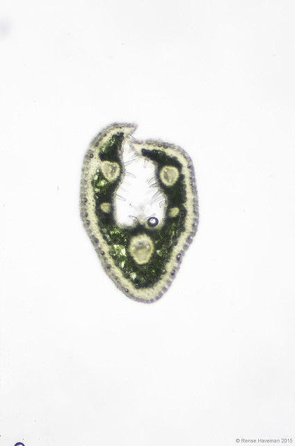
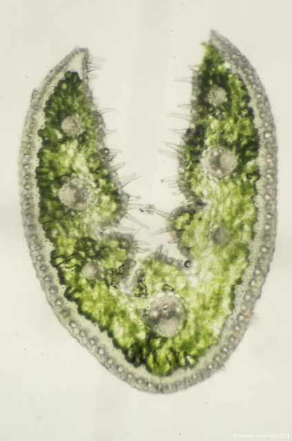
This weekend I experimented a bit with cross sections of fescue leafs. Sheep fescues are a notoriously difficult genus, and for the identification such cross sections are often needed. Important features of the anatomy of sheep fescues: the development of sclerenchyma (the light band at the outer side of the leaf), the number of veins and furrows, and the number and length of hairs at the inner side of the leafs.
Here three examples, Festuca filiformis, F. lemanii, and F. brevipila. All at 7x9 magnification.
What I learned: I have to clean the glasswork of the microscope very well, otherwise it's one spotted misery... Alcohol is your best friend in such microscope photography...

Festuca filiformis by Rense Haveman, on Flickr

Festuca lemanii by Rense Haveman, on Flickr

Festuca brevipila by Rense Haveman, on Flickr
This weekend I experimented a bit with cross sections of fescue leafs. Sheep fescues are a notoriously difficult genus, and for the identification such cross sections are often needed. Important features of the anatomy of sheep fescues: the development of sclerenchyma (the light band at the outer side of the leaf), the number of veins and furrows, and the number and length of hairs at the inner side of the leafs.
Here three examples, Festuca filiformis, F. lemanii, and F. brevipila. All at 7x9 magnification.
What I learned: I have to clean the glasswork of the microscope very well, otherwise it's one spotted misery... Alcohol is your best friend in such microscope photography...

Festuca filiformis by Rense Haveman, on Flickr

Festuca lemanii by Rense Haveman, on Flickr

Festuca brevipila by Rense Haveman, on Flickr

